Hand to Shoulder Center of Wisconsin’s orthopedic doctors and staff understand elbow fractures and elbow instability as well as the problems and pain these injuries can cause. Learn more about the causes of these injuries and how our orthopedic specialists treat them. Contact us today for more information.
Elbow Fracture vs. Elbow Instability
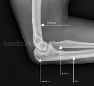
Figure 1: The three bones of the elbow
The elbow is a complex joint made up of three bones; the humerus, ulna, and radius (Fig. 1). The humerus is the large bone that extends from the shoulder to the elbow. The ulna and radius are located in the forearm; going from the elbow to the wrist. In addition to the three bones, the elbow also houses nerves, blood vessels, strong ligaments that hold the bones together, as well as muscles and tendons which help to maintain your elbow’s stability and guide movement of the joint.
An elbow fracture occurs when one or more of the bones are broken in the elbow. Elbow instability occurs when there is a loss of integrity to either the ligaments on the inside and/or outside of the elbow. This often happens with a fall on an outstretched hand when the elbow is dislocated. Sometimes, but not always, a dislocation is associated with a fracture. A fractured elbow typically occurs from a trauma however certain health conditions and diseases can increase the risk of a fracture. Loose bodies also commonly occur in the elbow as a result of small pieces of cartilage or bone floating within the joint. These loose bodies can cause the elbow to “catch” or “lock” at varying times.
Symptoms of a Fractured Elbow
Elbow injuries happen in both adults and children. The elbow is the most commonly dislocated joint in children. Common trauma and health conditions may cause instability and/or a fracture to the elbow.
Elbow fractured symptoms typically include severe pain, tenderness, difficulty in bending and straightening the elbow, and numbness in the fingers, hand or forearm. Additional visual symptoms such as a lump or deformity of the elbow, swelling and discoloration from bruising or redness may be present.
Elbow Fracture Types and Treatment
At Hand to Shoulder Center of Wisconsin, our sub-specialty trained orthopedic surgeons evaluate past and present medical history. A physical examination of the injured extremity is done and symptoms are discussed. Elbow x-rays are taken and evaluated. Depending on the degree of injury, a Computerized Tomography (CT) may be ordered. In rare cases, a Magnetic Resonance Imaging (MRI) may be ordered.
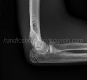
Figure 2: X-ray view of a child’s olecranon stress fracture
The radius bone is the smaller forearm bone that lies on the thumb-side of the forearm and connects with the upper arm bone (humerus) at the elbow. The radial head (or head of the radius) is located on the outer aspect of the elbow. The olecranon is located at the tip of the elbow. It connects and controls the powerful triceps’ muscle of the arm which straightens the elbow. The end of the humerus forms a spool-shape, which meets the ulna, allowing for elbow motion.
Because the elbow is made up of three bones, different types of elbow fractures can occur. Common fractures to the elbow include, radial head fracture, olecranon fracture (Fig. 2), and distal humerus fracture, also known as a supracondylar fracture.
Treatment for a fractured elbow vs. elbow instability varies greatly. Depending on the severity of the elbow instability, conservative treatment is often recommended. Physical or occupational therapy is ordered and if needed, a protective splint is provided to strengthen and stabilize the muscles around the joint. Rest and cutting back on strenuous activities along with bracing the elbow to keep it in a stabilized position is helpful. If symptoms do not improve overtime, surgery may be needed to repair or reconstruct the ligaments surrounding the elbow joint to restore stability.
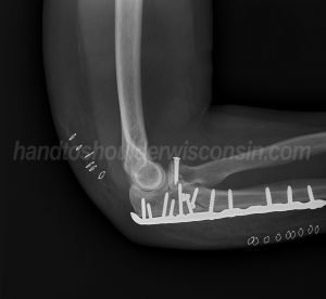
Figure 3: Severe fractures often require hardware to stabilize the bone and bone fragments
As for elbow fractures, certain fractures don’t require surgery; more traumatic fractures such as compound fractures and open fractures do require surgical intervention. Depending on the type of fracture(s), bone fragments, and the stability of the elbow joint, wires, plates, and screws may be used to stabilize the broken bone(s) (Fig. 3). All tendon, ligament, and/or blood vessel damage is also addressed during surgery.
Surgery for an elbow fracture is generally performed as an outpatient procedure. The surgeons at Hand to Shoulder Center of Wisconsin perform surgeries at Woodland Surgery Center and the three Fox Valley hospital locations. Following surgery, occupational or physical therapy sessions will be ordered to help regain range of movement and muscle strength. Therapists at Hand to Shoulder Center will focus on treating postoperative swelling, improving range of motion, improving strength, and restoring function.
On average, fractures take about six to eight weeks to heal, typically less for children. Total healing time is roughly three months. With or without surgery, the most common complication from an elbow injury is stiffness.